Overview
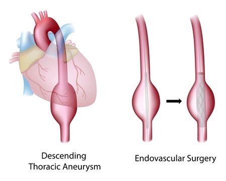
The aorta is a major blood vessel that delivers oxygen-rich blood from the heart to other organs and parts of the body. The aorta originates at the upper part of the heart but then turns downward and passes through the chest and the abdomen, producing branches on its way.Due to the central role of the aorta in the blood circulations, its failure to function properly has major negative consequences. If the wall of the aorta becomes weakened, the blood vessel starts to expand forming a bulge. This bulge is called aneurysm. If the bulge is located in the part of the aorta passing through the chest, it is referred to as thoracic aortic aneurysm or TAA.
Normal width of the aorta is 2 to 3 cm. At the site of an aneurysm, its diameter can expand to 5-7 cm and even more in some cases. Progressively bulging aorta has increasingly weakened wall. This wall can eventually rupture often causing death within minutes.

Symptoms
Thoracic aortic aneurysm can be asymptomatic. If, however, the expanding bulge starts to compress the surrounding tissues, a number of symptoms can be observed. They include chest or back pain, coughing, sneezing and difficulties with breathing and swallowing. The symptoms are not specific for aneurysm only and can relate to many different conditions.
Risk factors
Thoracic aortic aneurysm is much more common among males and usually observed in patients who are older than 50. It happens more often in people suffering from atherosclerosis and high cholesterol level. Smoking, excessive body weight, lack of physical activity, high blood pressure, family history of atherosclerosis or aneurysm and chronic obstructive pulmonary disease are some of the risk factors associated with the development of this life-threatening condition.
Diagnosis
A number of diagnostic tests can be performed when symptoms indicate the possibility of thoracic aortic aneurysm. Chest X-ray can show if the aorta is widened. Transthoracic echocardiography is an ultrasound investigation of the chest cavity. It also can show the presence and location of the aneurysm.The best diagnostic techniques are MRI scan and computer tomography (CT). They provide detailed image of the aorta and clearly show the size and exact position of the aneurysm.
It is recommended that people with increased risk developing this disease undergo regular checks aimed to detect any potential thoracic aortic aneurysm at early stage. If a small aneurysm is detected, further checks should be performed on a regular basis to monitor the aorta for signs of any negative developments.
Treatment
In cases when thoracic aortic aneurysm grows in size to more than 5-5.5 cm, expands very quickly (more than 1 cm per year) or becomes symptomatic (for example, causes breathing difficulties), the surgical treatment for this condition is usually recommended.There are three major types of surgery that can be performed: open chest surgery, keyhole surgery, and endovascular aneurysm repair. All three types of surgeries aim to place a graft at the site of the aneurysm. A graft is made of elastic synthetic material. Once the graft is inserted in the weakened area of the aorta, the blood starts flowing through it, thus taking away the pressure from the aneurysm and preventing it from getting bigger.

Open surgery involves opening of the chest, while keyhole surgery is done through several small cuts in the chest. Endovascular aneurysm repair is done with the use of stent-graft, a tube covered with graft material. The stent-graft is inserted through the femoral artery in the groin. Using X-ray images, the vascular surgeon guides the stent up to the site of the aneurysm in the chest, where it will bond to the wall of the aorta.
The choice of particular surgical technique depends on your health condition and the location and size of the aneurysm. The success of the treatment depends, in a significant degree, on the knowledge and experience of your doctors. Choosing the right specialists will help you to avoid many complications associated with the treatment of thoracic aortic aneurysm.


